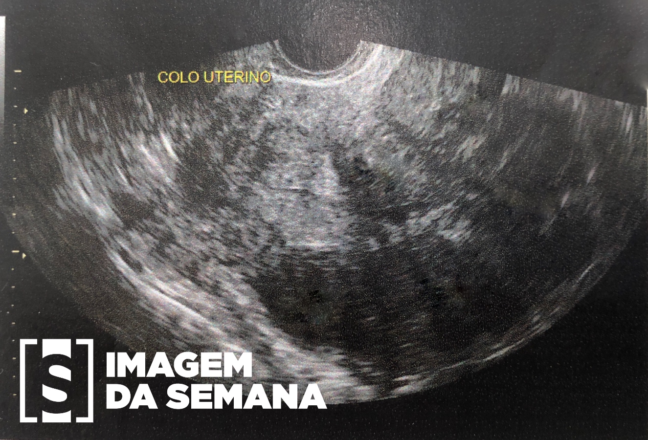
Case 200 Medical history Female patient, 34 years of age, never been pregnant, attended the gynecology service reporting infertility. Refers menarche at age 13, regular cycles and normal menstrual flow. Denies dyspareunia or pelvic pain. Presents hypothyroidism clinically controlled with levothyroxine. Physical examination and laboratory review unremarkable, except for the serum levels of CA-125: 91.2 IU / mL (Reference value: 35 IU / mL). Transvaginal ultrasonography (US) was requested (Image 1). Due to the findings, was prescribed the use of oral contraceptives (OC) for 6 months, however, there was no resolution of the infertility. Imagem 1: Ultrassonografia transvaginal em topografia de ovário esquerdo, evidenciando formação cística, ovalada, de paredes bem definidas, preenchida por ecos de baixa intensidade, medindo 3,8 x 3,0 x 3,5 cm – volume de 21,8cm3. Estudo com Doppler evidenciou fluxo restrito à periferia da estrutura referida.Image 1: Transvaginal Ultrasonography surveying the left ovary, showing a cystic, oval formation with well-defined walls, filled with low-level echoes, measuring 3.8 x 3.0 x 3.5 cm - Volume 21,8cm3. Doppler study showed flow restricted to the periphery of the structure. Question:Based on the clinical history and the imaging studies presented, which is the most likely diagnosis? Hemorrhagic luteal cyst Ovarian teratoma Mucinous cystadenoma Endometrioma Time is Up! Time's up