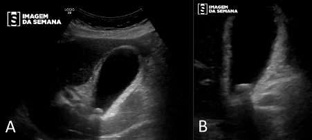
Medical history A 72-year-old female presented in the emergency room with right hypochondrium (RH) pain for the past 18 hours, initially with cramps and later continuously, accompanied by vomits. Previously diagnosed with arterial hypertension, type 2 diabetes and hypertriglyceridemia. No history of etilism. Good general condition, without jaundice and afebrile. Flat abdomen, flaccid, painful on light and deep palpation of the RH, interrupting deep inspiration during palpation. An upper abdominal ultrasound was ordered (images 1 and 2). Upper abdominal ultrasound, longitudinal plane demonstrating the gallbladder in left lateral decubitus (A) and dorsal decubitus (B). Question:Considering the clinical features and the images above, which diagnosis is the most likely? Acute cholecystitis Acute cholangitis Perforated duodenal ulcer Uncomplicated cholelithiasis Test question UNIVERSITARY HOSPITAL PRESIDENTE DUTRA - MA (2016). A 40-year-old woman, obese, multiparous, with history of epigastric pain radiating to righ hypocondrium for the past 2 years, associated with nausea, vomiting and isolated episodes of fever. Went through abdominal ultrasound 1 year ago, which evidenced cholelithiasis (with small and medium calculus). Reporting upper abdominal pain started 3 days ago, radiating to the back, choluria, jaundice, fever and chills. Submitted to cholangio resonance, which evidenced cholelithiasis, choledocholithiasis (3 bile duct calculus) and choleduchus 14mm dilated.Question:Regarding the written case, point out the INCORRECT alternative. Female gender, age, obesity, multiparity can be considered risk factors to biliary lithiasis Secondary biliary lithiasis can be considered the most common cause of cholelithiasis, and the gold standard treatment in symptomatic patients is cholecystectomy The patient presents with clinical criteria to the Charcot triad, that might be associated with acute cholangitis, which might demand decompression of the main biliary tract The use of systemic antibiotics for enteric gram-negative germs is justified, besides the control of the infection focus Because of the risk of bleeding, duodenal perforation and acute pancreatitis, retrograde endoscopic cholangiopancreatography with papillotomy and choledocolitotomy is not recommended Time is Up! Time's up