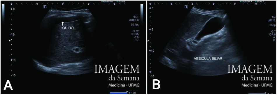
Enunciado 83 year-old male patient, admitted at the Emergency Room with right hypochondrium intense pain. Physical examination reveals painless hepatomegaly, without further alterations. Murphy and Courvoisier signs were both negative. Personal history of chronic kidney disease stage 4, hypertension and abdominal aorta prothesis. Social drinker, former smoker, denies liver diseases or family history of those. Liver enzymes altered, anaemia and alpha-fetoprotein within normal range. Imaging exams are shown as requested. Upper abdomen ultrasound. Liver, cross sectional (A) and gallbladder, longitudinal sectional (B). Upper abdomen ultrasound. Liver, right lobe, B-mode study (1a, 2a, 3a) and Power Doppler (1b, 2b, 3b). Abdominopelvic magnetic resonance imaging (MRI), T2-weighted, no contrast. Axial view of the upper abdomen (A) and abdominopelvic coronal view (B). Abdominopelvic multislice computed tomography (CT), no contrast. Axial view of upper abdomen (A) and abdominopelvic coronal view (B). Question:Considering the clinical data and the images, the most adequate conduct to better elucidate the diagnosis is: Percutaneous core needle biopsy Laparotomy with right lobectomy Contrast enhanced magnetic resonance imaging Contrast enhanced ultrasound Time is Up! Time's up