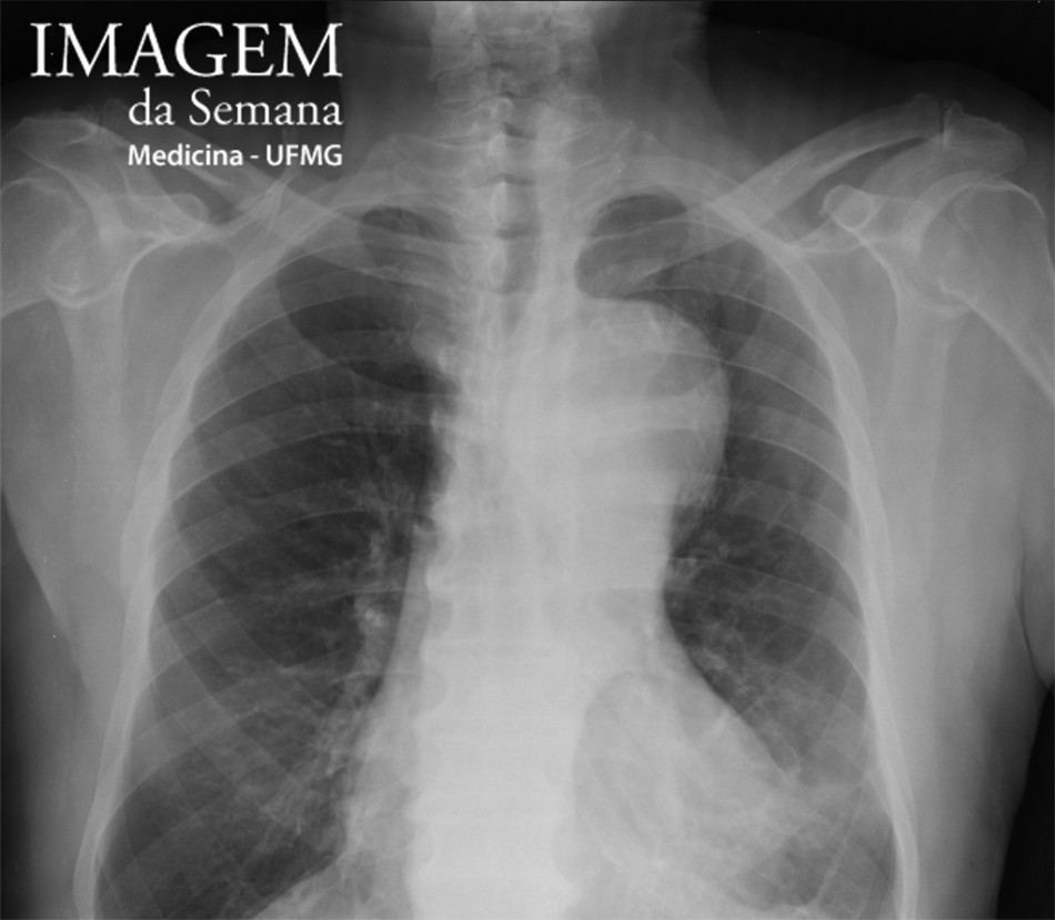
Medical history Male patient, 71 years old, referred due to complaint of upper abdominal pain with radiation to the back in three episodes in the last three months. There are not aggravating or relieving factors, as well as episodes of subtle worsening of the pain. Hypertensive, former smoker (ceased 20 years ago). Previous CVA report, without significant sequelae and without history of thoracic trauma. Submitted to radiological propedeutics, including chest radiography and, subsequently, thoracic and abdominal CT angiography. Image 1: Chest radiograph, posteroanterior view, orthostatic position. Image 2: Chest radiograph, lateral view, orthostatic position. Image 3: Thoracic and abdominal aorta CT angiography, sequential coronal reconstructions. Image 4: Thoracic aorta computed tomography (CT) angiography, axial view, at the aortic arch level, before (a and b) and after iodinated intravenous contrast injection, arterial phase (c and d). Question:Considering the clinical history and the presented images, which one is the most likely diagnosis? A) Dissecting aneurysm of the thoracic aorta B) Thoracic aortic pseudoaneurysm C) Saccular aneurysm of the thoracic aorta D) Fusiform aneurysm of the thoracic aorta Test question (Unifesp – 2015) 22 years old patient with Marfan syndrome (MS), asymptomatic, with family history of MS, in semiannual evaluation, currently presents ascending thoracic aorta aneurysm of 5,5 cm. Choose the most appropriate option.. a) Continue the follow-up every six months b) Increase the dose of the beta blocker c) Surgery must be indicated d) Surgery must be indicated only if the aortic diameter is > 6,0 cm e) Surgery does not change the natural history of the Marfan syndrome Time is Up! Time's up