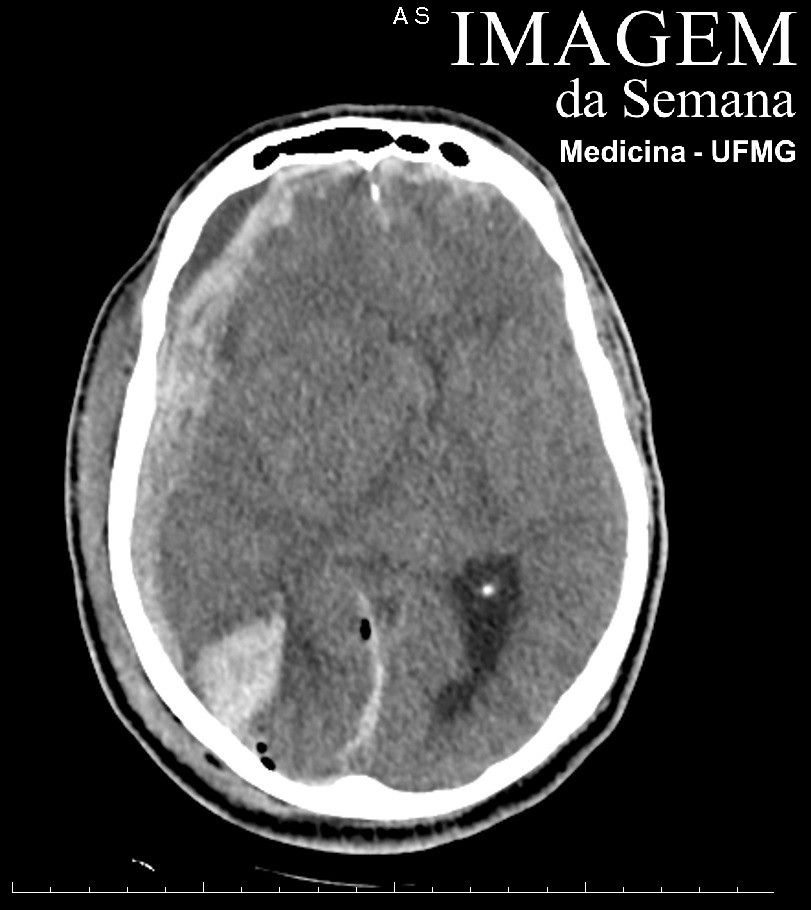
Medical history A 66 year-old male patient is admitted unconscious to a trauma center after falling from a 3 meter height. His primary examination reveals: patent airway, symetric thoracic expansibility, non-laborious breathing, normal breath sounds, 22 respiratory incursions per minute, hemodynamic stability, 8 points in the Glasgow Coma Scale (GCS), isocoric non-photoreactive pupils and right otorrhagia. A non-contrast-enhanced head computed tomography (CT) scan is urgently acquired. Image 1: Non-contrast-enhanced head computed tomography (CT), axial section, level of the occipital horns of the lateral ventricles, brain window. Image 2: Non-contrast-enhanced head computed tomography (CT), axial section, level of the frontal horns of the lateral ventricles, brain window. Image 3: Non-contrast-enhanced head computed tomography (CT), coronal reconstruction, level of the mastoid parts of the temporal bones, bone window. Question: Taking into account the clinical scenario and the CT head scan results, what are the next best steps in this patients management? a) Orotracheal intubation and neurosurgery b) 12 to 15 L/min oxygen therapy delivered by a mask-reservoir device and neurosurgery c) Orotracheal intubation, 20% manitol and close monitoring in the next 24 hours d) Orotracheal intubation, agressive fluid resuscitation and close monitoring in the next 24 hours None Test question [Medical Residency 2017: Faculty of Medical Sciences of Unicamp – SP] A 7 year-old male patient is brought unconscious and with spontaneous breathing to the Emergency Department by Emergency Medical Services after having been run over and thrown a distance of approximately 10 meters. Physical examination: HR = 140 bpm; RR = 33 ripm; capillary refill time = 3 seconds. Head: frontal contusion. Neurological examination: the patient emits incomprehensible sounds, localizes pain and doest not open his eyes in response to painful stimuli. Question:The patient’s conscious state and the best next step in airway management are: a) Glasgow Coma Scale = 8; orotracheal intubation b) Glasgow Coma Scale = 11; mask-reservoir device c) Glasgow Coma Scale = 9; nasal cannula with an oxygen flow of 5 L/min d) Glasgow Coma Scale = 6; laryngeal mask airway None Time's up