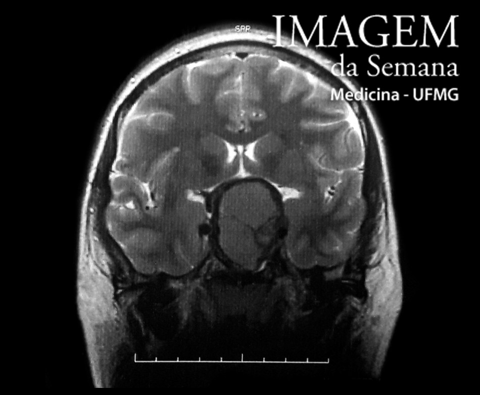
Medica history A 33-year-old male patient seeks out eye care for loss of progressive visual acuity in the temporal visual field of the right eye for 15 days. He refers episodes of left hemicranial headache which started four years ago as well as loss of libido. Laboratory tests revealed TSH: 1,850 μUI / mL (RV: 0,465 to 4,680), total testosterone: 92 ng / dL (RV: 132 to 813), prolactin: 7111 ng / mL (RV: 9), FSH: 2.25 mIU / mL (RV: 1.55 to 9.74), LH: 1.97 mIU / mL (RV: 0.82 to 6.22) and baseline cortisol: 12.3 μg / dL (RV: 4.46 to 22.7) and magnetic resonance imaging (MRI) of the brain (presented). Magnetic resonance imaging of the brain, T2-weighted, coronal cut, sellar and suprasellar level, with no intravenous injection of paramagnetic contrast agent (gadolinium). Magnetic resonance imaging of the brain, T2-weighted, coronal cut, suprasellar level, with no intravenous injection of paramagnetic contrast medium (gadolinium). Magnetic resonance imaging of the brain, T1-weighted, axial cut, suprasellar level, after intravenous injection of paramagnetic contrast agent (gadolinium). Question:Considering the clinical history and the complementary propaedeutic results, which is the most likely diagnosis? Craniopharyngioma Prolactinoma Meningioma of Sella Turcica Pituitary metastasis Test question Question:(Medical residency - UFPR - 2008) What is the most frequent pituitary adenoma and which is the most used initial therapeutic orientation? a) GH secreting adenoma - radiosurgery b) Non-functioning adenoma - surgery and radiation therapy c) Prolactin secreting adenoma - dopaminergic agonist d) Prolactinoma - transsphenoidal surgery e) ACTH secreting adenoma - ketoconazole Time is Up! Time's up