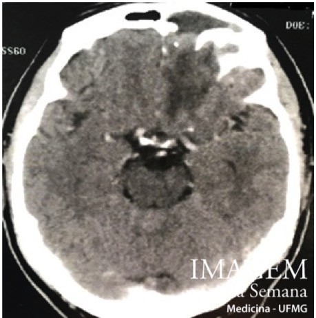
Enunciado A 57 year-old male patient, with serous rhinorrhea, nasal congestion , dry cough and sneezing for 30 days developed nasal discharge, purulent expectoration, sensation of pressure in the frontal region, severe headache and fever, with no meningeal signs upon physical examination. He was diagnosed with frontal sinusitis and treated with levofloxacin, without conducting additional workup. On the 9th day of antibiotic therapy, had no response to the established treatment and evolved with episodes of seizures. A computed tomography (CT) and magnetic resonance imaging (MRI ) of the brain were requested. Image 1: Computed tomography of the skull, axial view, brain-window. Image 2: Magnetic Resonance Imaging of the skull, axial view, weighed in T2, at the level of the thalamus. Image 3: Magnetic Resonance Imaging of the skull, axial view, diffusion weighed, at supraventricular level. Question:Considering the clinical history and images, which is the most likely diagnosis? Bacterial meningitis Frontal sinus osteoma Brain abscess Thrombosis of venous sinuses Time is Up! Time's up