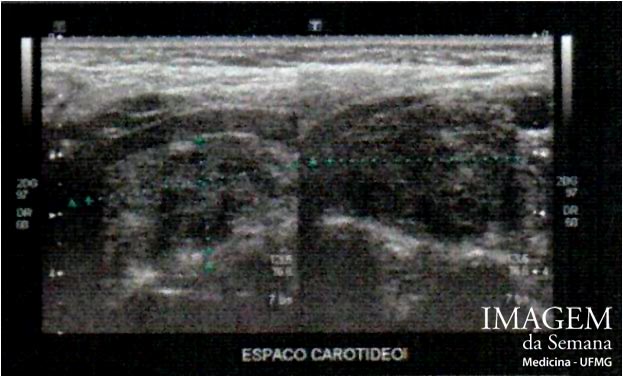
Medical history A 44-year-old female patient attended a medical appointment due to a lump she had recently noticed in the upper third of her neck, on the right. At clinical examination, a firm, painless and pulsatile mass was felt in this area. It could be moved more easily side-to-side than up and down. Besides, it was observed a bulge in the right lateral wall of her oropharynx extending to the midline. Figure 1: Neck ultrasonography – focus on the carotid spaceFigure 2: Neck ultrasonography – focus on the carotid spaceFigure 3: Computed tomography of the neck - after injecting contrastFigure 4: Computed tomography of the neck - three-dimensional reconstruction Question:Based on the clinical data and imaging studies, which is the most likely diagnosis? Neck schwannoma (neurinoma) Parotid pleomorphic adenoma Internal carotid artery aneurysm Carotid body paraganglioma None Time's up