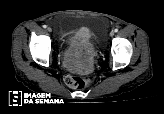
Medical history A 56-years-old G3Pv3A0 woman that went into menopause 5 years ago presents to the emergency department with intense pelvic pain for 1 year that got worse in the past 4 days. History of moderate vaginal bleeding and constipation, requiring enema for evacuation. She lost 44 lbs in the past 6 months. Tobacco and alcohol user. The speculum examination reveals friable cervix with an exophytic lesion. Bimanual examination reveals firmness on the vaginal wall and uterine cervix. With the hypothesis of cervical cancer, computed tomography and anatomopathological examination of a sample were performed. Intravenous contrast-enhanced computed tomography of the pelvis, axial view, section level of the bladder, portal venous phase.Intravenous contrast-enhanced computed tomography of the pelvis, axial view, section level of the uterine corpus, portal venous phase.(A) Intravenous contrast-enhanced computed tomography of the abdomen, axial view, section level of the kidneys, portal venous phase.(B) Intravenous contrast-enhanced computed tomography of the abdomen, axial view, section level of the renal pelvis, portal venous phase. Question:By analyzing the images, which important finding for staging is NOT found? Bladder invasion Colonic invasion Lymph node metastases Hydronephrosis Test question (Hospital das Clínicas de Porto Alegre – 2015) 45 years old woman attended medical appointement due to intermitent genital bleeding. She was using oral contraceptive and the last gynaecological examination was dated more than 5 years before the present appointment. At examination, it was visualized a 5 cm lesion emerging from the cervix and reaching the upper third of the vagina. Rectal examination revealed parametrious thickening, without pelvic fixation. Biopsy showed epidermoid carcinoma.Question:What is the most recommended treatment? Radiotherapy and chemotherapy Wertheim-Meigs surgery Pan-hysterectomy and bilateral oophorectomy Total hysterectomy Conization Time is Up! Time's up