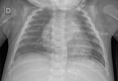
Enunciado A 3 months old male patient, asymptomatic, undergoes investigation of hepatomegaly observed in the physical exam and confirmed by abdominal ultrasound showing multiple liver nodules. There is no report of jaundice or family history for liver diseases. Medical discharge at birth with mother and good weight gain. On physical examination, he presented a cutaneous nodule in the right hypochondrium. Requested propaedeutic imaging (images 1 to 4). Image 1: Posteroanterior chest radiograph. Image 2: Computed tomography (CT) of the chest, abdomen and pelvis, with iodinated contrast medium. Sagittal (A), coronal (B) and axial (C) cuts. Image 3: Magnetic resonance (MRI) of the thoracic spine, left paramedian sagittal section T1-weighted image (A), left paramedian sagittal section T2-weighted image (B), and middle sagittal section T2-weighted image (C). Image 4: Computed tomography (CT) of the chest, abdomen and pelvis, with iodinated contrast, coronal cut (A) and axial cuts (B and C). Based on the clinical history and the images, which is the most likely diagnosys? Hepatoblastoma Thoracic Lymphoma Neuroblastoma Germ Cell Tumor Questão de Prova [BRAZILIAN SOCIETY OF PEDIATRICS – Title of Pediatrician specialist] Four-year-old female, with a history of fever for a month, is taken to the clinic. Physical examination: hypocolored + / 4. Laboratory tests: hematocrit: 29%, ESR: 85 mm / 1st hour, MCV: 85fl. Chest X-ray: massive posterior mediastinal mass of irregular contours with calcifications. This clinical condition is suggestive of: Lymphoma Pneumonia Neuroblastoma Bronchogenic cyst Ganglionic Tuberculosis Time is Up! Time's up