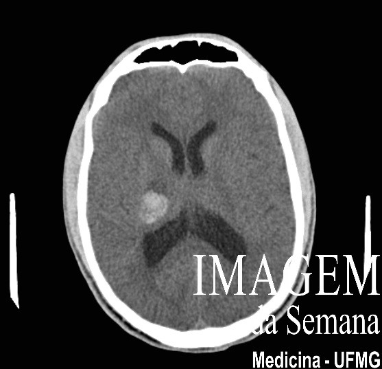
Medical history 19 year-old male patient refers 2 days with difficulty to walk and slightly ataxic gait. History of neuropsychomotor developmental delay, with difficulty to learn in early childhood. Denies comorbidities. Attending doctor requested a brain CT (Image 1) which revealed lesion in the right internal capsule area. Patient also brought previous MRI results (Image 2 and 3). Image 1: Computed tomography (CT) of the brain, axial view, lateral ventricles level, without injection of intravenous iodine-based contrast. Image 2: Magnetic resonance imaging (MRI) of the brain, T2-weighted, axial view, lateral ventricles level. Image 3: Magnetic resonance imaging (MRI) of the brain, gradient-echo, axial view, lateral ventricles level. Question:Considering the information provided and the images shown, the probable aetiology for this case is: A) Developmental venous anomalies B) Arteriovenous malformation C) Hemangioblastoma D) Cavernoma Test question (Rio de Janeiro Military Police, Neurosurgery – 2010) The lesion most frequently associated to cavernoma is: a) Basilar aneurysm b) Capillary telangiectasia c) Venous angioma d) Dural arteriovenous malformation Time is Up! Time's up