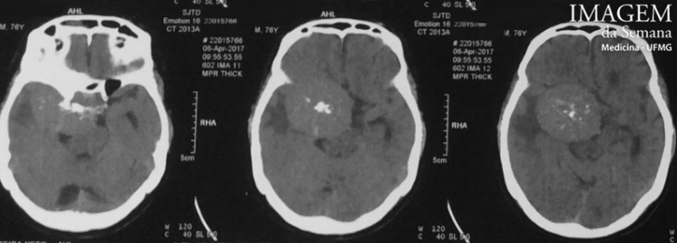
Medical history A 76-year-old male patient was taken by family members to medical care at a Basic Health Unit with an intermittent memory alteration (possibly lost in the peridomiciliary environment) starting 2 years ago. In addition, he complained of progressive deterioration of gait and episodic headache, both with 1 year of evolution. He is hypertensive with inadequate control. Referred to HC-UFMG for hospitalization, after the accomplishment of computed tomography (CT) of the encephalon. Image 1: CT scan of the brain, axial view at the sealing/supraselar level, without intravenous contrast. Image 2: CT scan of the brain, axial view at the suprasellar level, after intravenous injection of iodinated contrast. Image 3: Magnetic resonance imaging (MRI) in T2 sequence, axial view at the suprasellar level. Image 4: Magnetic resonance imaging (MRI) in T1 sequence after administration of intravenous contrast (gadolinium), sagittal view at the right parasselar level. Question:Based on the clinical history and the presented images, the most probable diagnosis is: A) Glioblastoma multiforme B) Metastasis dural C) Meningioma D) Arachnoid cyst Test question (HC – UFPR)(Specific Test / 2016) Meningiomas are usually benign tumors of slow growth. The most common location of intracranial meningiomas in adults is: a) Parassagital b) In the convexity c) Intraventricular d) In the olfactory groove e) In the cavernous sinus Time is Up! Time's up