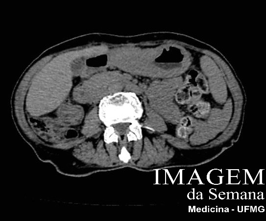
Medical History 71 year-old female patient, former smoker, refers postprandial fullness, early satiety, weight loss (15kg in 5 months) and adynamia. Physical examination reveals emaciation, pallor and a palpable epigastric mass, hardened and mobile, with 6cm in diameter. Upper gastrointestinal (UGI) endoscopy showed ulcero-infiltrative lesion, with 5cm in extension, occupying the whole circumference of stomach's distal-third. Histology was inconclusive for gastric cancer. Abdominopelvic computerized tomography (CT) was requested, as shown. Abdominal computerized tomography, axial plane, without iodine-based intravenous (IV) contrast, at the level of gastric antrum and body.Abdominal computerized tomography, axial plane, after injecting iodine-based IV contrast, at the level of gastric antrum and body. Abdominopelvic computerized tomography, coronal reconstruction, after injecting iodine-based IV contrast, at the level of gastric antrum and body.Question:After analyzing the clinical data and the images, which one is the most likely diagnosis? Gastric large B-cell lymphoma. Diffuse type gastric adenocarcinoma (Lauren classification). Intestinal type gastric adenocarcinoma (Lauren classification). Corrosive antritis. Test Question (Medical Residence 2017 - Universidade Federal de São Paulo) 65 year-old female, referring epigastric pain for 3 months, performed an UGI endoscopy which found ulcerative lesion with 2,2cm in gastric fundus. Histologic report confirmed indifference gastric adenocarcinoma, with signet-ring cells. CT showed just gastric lesion, without metastasis. Question:Patient should be submitted to: Total gastrectomy with D2 lymphadenectomy. Neoadjuvant chemoradiotherapy. Endoscopic mucosectomy. Subtotal gastrectomy with D2 lymphadenectomy. None. Time is Up! Time's up