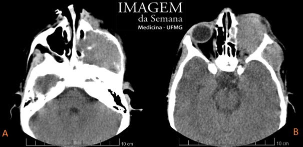
Medical History 5-year-old male patient presents with left facial tumefaction and ipsilateral ptosis for the last 15 days. He denies fever, pain, headache or head trauma. His physical examination is remarkable for left ptosis, left orbital tumefaction with frequent eye tearing, but neither skin nor conjunctival hyperemia. Mouth deviation to the right, tongue deviation to the left and severe left visual acuity loss (capable of seeing only shadows). Non-contrast-enhanced head computed tomography (CT), axial sections at the level of the maxillary sinuses (A) and orbits (B). Contrast-enhanced head computed tomography (CT), axial sections at the level of the maxillary sinuses (A) and orbits (B). Contrast-enhanced T1-weighted head magnetic resonance imaging (MRI), axial section at the level of the maxillary sinuses and nasal cavity. Contrast-enhanced T1-weighted head magnetic resonance imaging (MRI), coronal section at the level of the maxillary sinuses and orbits. Pergunta:Based on the clinical case and the images provided, what is the most likely diagnosis? Orbital cellulitis. Burkitt's lymphoma. Neuroblastoma. Rhabdomyosarcoma. Test Question (HUPES – UFBA – Pediatric Oncology – 2014) Concerning rhabdomyosarcomas, select the correct alternative: They are benign tumors that more frequently affect children under three years old. They originate from skeletal striated muscle cells. Rhabdomyosarcomas are notable for the fact that they always present with important inflammatory signs in the neighboring areas of the tumor. Histologically, the embryonal subtype is the rarest. Children under one year old have the best prgnosis, independent of other factors. Time is Up! Time's up