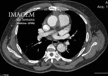
Medical History A 56- year-old man attends the emergency department complaining of severe, acute, breathing-related chest pain radiating to the abdomen. Long-standing history of controlled hypertension, in use of Beta-Blocker. On examination: patient in pain, pallor, sweating. No abnormalities were observed in the respiratory, cardiovascular and digestive systems. BP: 155/94 mmHg; HR; 88 bpm; RR: 22 bpm; SpO2: 96%. A computed tomographic (CT) angiography of the abdominal and thoracic aorta was requested. Image 1: CT angiography of the aorta, axial section, at level of pulmonary arteries, mediastinal window. Image 2: CT angiography of the aorta, coronal reconstruction, at level of descending aorta, mediastinal window. Image 3: CT angiography of the aorta, sagittal reconstruction, at level of descending aorta, mediastinal window. Question:Considering the images and the clinical data, which is the most likely diagnosis? a) Acute mesenteric ischemia b) Acute coronary syndrome c) Acute aortic dissection d) Pulmonary thromboembolism Time is Up! Time's up