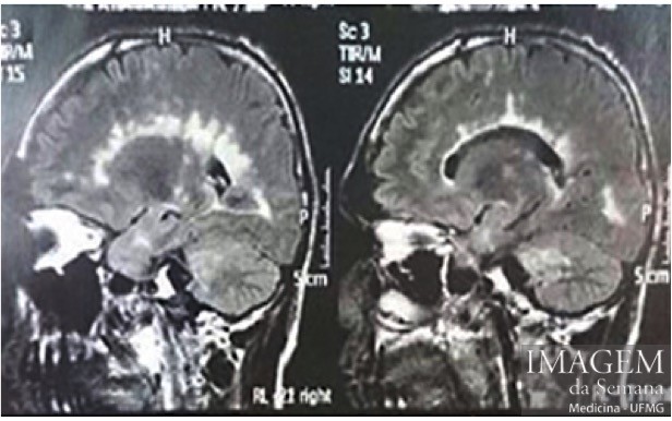
Medical History 32 years old, male patient presents a change in the walking patterns, motor coordination, vision and the speech, of slow progression for 14 years. At the time of the first symptoms, he lost control of the anal sphincters and he is in a wheel chair for 3 years. He denies acute symptoms or spontaneous improvement. At the examination, he has loss of the visual acuity, change in the postural kinetic sensitivity, presence of the Babinsky sign and bilateral hyperreflexia of the Achilles tendon, and global muscular strength reduced in the lower limbs. Image 1: Flair-Weighted Magnetic Resonance imaging of the cranium, Sagittal plane. Image 2: T1-weighted MR imaging of the cranium, Axial plane. T2-weighted MR imaging of the cranium, Axial plane. Question:Based on the clinical history and in the imaging studies presented, what is the most likely diagnosis? a) Multiple sclerosis b) Wilson's disease c) Lateral Amyotrophic Sclerosis d) Machado–Joseph disease Time is Up! Time's up