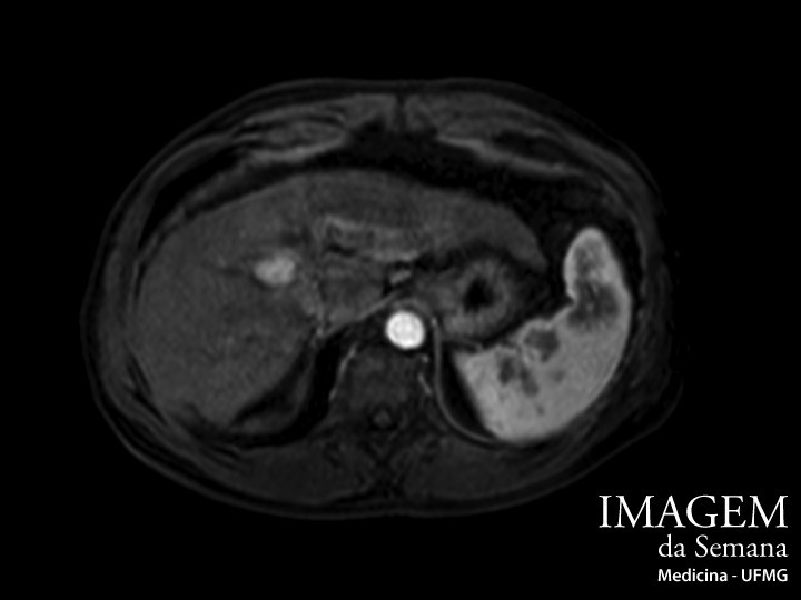
Enunciado Male patient, 61 years-old, diagnosed with hepatitis C 23 years ago. Currently has cirrhosis with mild portal hypertension and small-caliber esophageal varices, but remains well adjusted (Child-Pugh score A5). In the ultrassound screening, a solid hyperechoid nodule between segments VIII and IV of the liver was identified. A magnetic resonance imaging of the abdomen with the injection of paramagnetic contrast was requested to better identify the lesion (Images attached). Image 1: Magnetic Resonance Imaging (MRI) of the abdomen, weighted in T1, after venous contrast, arterial phase. Image 2: Magnetic Resonance Imaging (MRI) of the abdomen, weighted in T1, after venous contrast, portal phase. Question:Taking into consideration the images and the patient data, what is the most likely diagnosis of the lesion? Hepatocellular carcinoma Metastatic tumor Hepatic hemangioma Cholangiocarcinoma Time is Up! Time's up