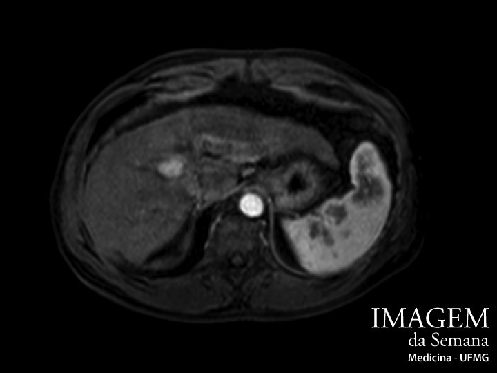
Enunciado Male patient, 61 years-old, diagnosed with hepatitis C 23 years ago. Currently has cirrhosis with mild portal hypertension and small-caliber esophageal varices, but remains well adjusted (Child-Pugh score A5). In the ultrassound screening, a solid hyperechoid nodule between segments VIII and IV of the liver was identified. A magnetic resonance imaging of the abdomen with the injection of paramagnetic contrast was requested to better identify the lesion (Images attached). Image 1: Magnetic Resonance Imaging (MRI) of the abdomen, weighted in T1, after venous contrast, arterial phase. Image 2: Magnetic Resonance Imaging (MRI) of the abdomen, weighted in T1, after venous contrast, portal phase. Question:Taking into consideration the images and the patient data, what is the most likely diagnosis of the lesion? Hepatocellular carcinoma Metastatic tumor Hepatic hemangioma Cholangiocarcinoma Anterior Próximo Próximo Time is Up! Time's up