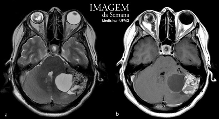
Enunciado Female patient, 44 years old, admitted with a recent case of ataxia and progressive holocranial headache, as well as reduced visual acuity in the left eye. Diagnosed with hypertension of difficult control, non-insulin dependent diabetes and amaurosis on the right eye (due to traumatic retinal detachment). Magnetic resonance imaging of the skull (image 1) and ophthalmoscopy of the left eye were requested and suggested a single etiology and guided the request for additional workup. (Images 2 to 4). Magnetic resonance imaging (MRI) of the skull, axial view, T2-weighted (a) and T1-weighted, after administration of a paramagnetic contrast agent, gadolinium (b). Abdominal computed tomography (CT), axial view, arterial phase, after administration of iodine contrast. Magnetic resonance imaging (MRI) of the abdomen, axial view, weighted in T2. Full-body scintigraphic study performed 24 and 48 hours after administration of 5 mCi of 131 Iodine-MIBG. Below, in black background, additional images with SPECT / CT technique in coronal, sagittal and transverse axes. - 1st line: CT images; 2nd line: SPECT scintigraphy images fused with the CT shown in the first line. Question:Taking into account the clinical presentation and the exams provided, what was the diagnostic hypothesis raised by the assistant team? Polycystic kidney disease Von Hippel-Lindau disease Tuberous Sclerosis Multiple Endocrine Neoplasia type 2 Time is Up! Time's up